Vitreoretinal Diseases
Vitreoretinal Diseases
Information regarding the most common diseases we treat can be found below.
Most patients are initially unfamiliar with vitreoretinal diseases and usually present to a retina clinic after learning that glasses will not help their vision in the immediate future. Understandably, this can be anxiety provoking for many of our patients. Please keep in mind that the information found below is for educational purposes. Nothing should substitute an evaluation by our Physicians.
Eye Anatomy

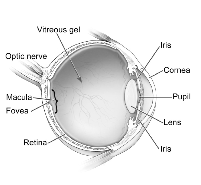
Parts of the Eye
- Vitreous Gel: A clear gel that fills the inside of the eye.
- Optic Nerve: A bundle of more than one million nerve fibers that carries visual messages from the retina to the brain.
- Macula: The small sensitive area of the retina that gives central vision. It is located in the center of the retina and contains the fovea.
- Fovea: The center of the macula and gives the sharpest vision.
- Retina: The light-sensitive tissue lining at the back of the eye. The retina converts light into electrical impulses that are sent to the brain through the optic nerve.
- Iris: The colored part of the eye that regulates the amount of light entering the eye.
- Lens: A clear part of the eye behind the iris that helps to focus light, or an image, on the retina.
- Pupil: The opening at the center of the iris. The iris adjusts the size of the pupil and controls the amount of light that can enter the eye.
- Cornea: The clear outer part of the eye’s focusing system located at the front of the eye.

